Positron Emission Mammography (PEM)
Naviscan Solo II
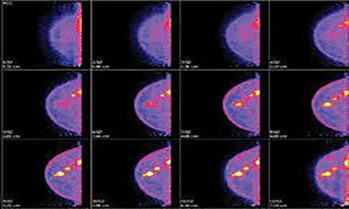
The Breast PET scanner (also called positron emission mammography (PEM)) produces high-resolution images, with far more detail than conventional breast imaging. By producing tomographic images of lesions with resolution down to 1.6 mm (approximately the size of a grain of rice), this technology provides valuable clinical data on invasive and non-invasive disease across the continuum of care. From initial staging to ongoing post-surgical disease management, CMR Naviscan’s high-resolution Breast PET Scanner provides a metabolic perspective allowing physicians to provide optimal patient outcomes.
CMR Naviscan offers a suite of products to aid in the detection and monitoring of breast cancer. The Naviscan Solo II™ High Resolution Breast PET Scanner System is a recognized problem-solving tool for complex cases and a valuable aid in treatment monitoring and/or surgical planning for pre and post treatment patients. Biopsies can then be performed same-day with our Stereo Navigator™, a secure, PET-guided accessory that obtains tissue samples of suspicious tumors. Finally, the MIMViewer® is an integrated platform for Breast PET viewing and analysis.
FEATURES
- Highest sensitivity and specificity in characterizing the extent and location(s) of cancer
- Precise lesion targeting through 3-D tomographic imaging and automated software
- High 1.6 mm spatial resolution
- Unlike mammography, Breast PET gently steadies the breast without compression
- Patient can sit comfortably and undergo minimal scan times with no claustrophobia issues
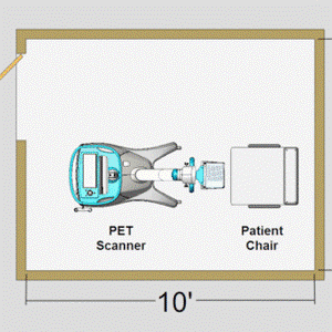
- Compact, portable, easy to use
Stereo Navigator – Software
The Stereo Navigator™ enables physicians to target and guide biopsy devices toward abnormalities visible on Breast PET, facilitated by the biopsy software that displays needle target position and suggested trajectory to obtain tissue samples for pathologic evaluation. The cutting edge in targeted molecular imaging of breasts, this PET-guided biopsy accessory is indicated for the localization of lesions in female breasts.
FEATURES
- Greater patient comfort and reduced anxiety with no claustrophobia or prone positioning issues
- Secure needle guide support for ease in device placement
- Precise lesion targeting in three dimensions through tomographic imaging and automated software
- Prompt verification of device positioning by line source scanning and software overlay
- Immediate confirmation of sampling accuracy with post-biopsy imaging
- Compatible with the following breast biopsy devices: Mammotome® Breast Biopsy System (Devicor™ Medical Products, Inc.), ATEC® Breast Biopsy System (Hologic, Inc.) and EnCor® Breast Biopsy System (C.R. Bard, Inc.)
MIMviewer
The MIMviewer’s customizable clinical reading workflows provide you with an automated and reproducible approach to image interpretation and analysis. A workflow, also known as a macro, is a sequence of scripted actions that are performed automatically by the software. Most of the major tools in MIMviewer can be added to workflows. You are able to register any number of studies automatically, seamlessly switch between multiple layouts, change color tables, and create screen captures, all without having to click a mouse. MIM provides an easy graphical interface to create these workflows, allowing you to adapt them to meet your specific needs. It makes the analysis of fusion images a simple task. The MIMviewer is from the family of MIM Software image viewing and analysis platforms
FEATURES
MIM can fuse any number of exams from multiple modalities, including CT, MR, PET, SPECT, CBCT, and more
Auto-alignment allows for the fast and accurate fusions and key to correlating any number of exams over time
Universal, secure (HIPPA compliant) access to imaging data from anywhere
MIM also provides tools for treatment monitoring using various response criteria such as RECIST and PT.ERCIS


Gamma Detection System (Gamma Probe)
The Navigator provides the best gamma probe value in the market. The Navigator 2.0 combines the reliability and features of its predecessor, the Navigator GPS, with state-of-the-art wireless probe technology in a new, sleek design. Lightweight and compact, this system is the ultimate in portability and the advanced battery technology allows for optimal placement of the control unit in the OR without the limitation associated with power cords. Unlike other systems, no calibration from probe to control unit is required before use; the system is ready to use the moment it is turned on.
KEY FEATURES:
- 100% Wireless – Industry Standard Technology
- Exceptional Accuracy – No Signal Delay
- Instant Power “ON” – No Probe Tethering or Calibration Required
- Extended Battery Life – 10+ Hours of Continuous Use
- Sterilizable Probes with All Major OR Sterilizers
The Navigator system is used in radio-guided surgical procedures, primarily for lymphatic mapping and tumor localization. Radio-guided surgical techniques using radiopharmaceuticals to locate a number of different tumor sites have been effective in the localization of other diseases, such as parathyroid adenomas and recurrent cancer. Most recently, MIS (Minimally Invasive Surgical) procedures, such as VATS, have proven to be effective in the localization of small occult tumors in the lung. This procedure allows for surgeons to stage cancer much earlier than the “wait-and-see” approach.

Select the various hotspots on the image below to learn how the Navigator 2.0 Wireless probes provide instant information during surgical procedures